About Us
Centers & Services
Conditions & Treatments
Make an Appointment
Fee Schedule / Budget Estimation
Patient Information
Find a Doctor
Insurance & Direct Billing
Facilities
Promotions
Health Guides
News & Seminars
Contact Us
2
EN
繁體
简体
日本語
2
EN
繁體
简体
日本語
Visiting Arrangements
Hospital Services During Bad Weather
View All
Urgent Care Center
Next Ticket Number for Stable Cases
Existing Consultation
Stable Cases Waiting for Consultation
Consultation will be given according to triage priority - Critical / Urgent
Center and Services
Facilities
Conditions & Treatments
Make an Appointment
Fee Schedule
Patient Information
Promotions
View More
Adventist Children's Health Hotline
1 Jun 2026
Father's Day Physical Check up Promotion
1 Jun 2026
Health Assessment Promotion, Physical Examination Services
Hospital Anniversary and Mother's Day Breast Screening Promotion
6 May 2026
Health Assessment Promotion, Physical Examination Services
Cardiac Assessment Promotion
3 May 2026
Cardiac Promotion
Health Guides
View More
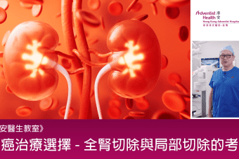
Consultant In Urology Dr. Chan Lung Wai: Early Detection of Kidney Cancer — Surgical Choice Matters for Preserving Kidney Function!
13 May 2026
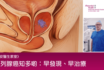
Consultant In Urology Dr. Chan Lung Wai: Men’s Health Alert! Screening and Treatment for Prostate Cancer
10 May 2026
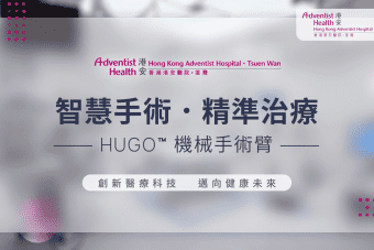
Hong Kong Adventist Hospital – Tsuen Wan introduces the Hugo robotic-assisted surgery system, ushering in a new era of intelligent minimally invasive surgery!
1 May 2026
Adventist Health Education Series — Must-Know During Pregnancy! Top 10 Pregnancy Tips to Help Expecting Moms Navigate the Critical Period with Peace of Mind!
1 Jan 2026
Adventist Health Education Series — Don’t Panic When Your Child Has a Fever! Must-Know Observation and Cooling Steps for Parents
21 Nov 2025
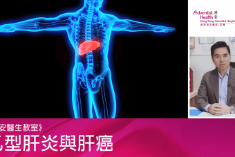
Adventist Health Education Series – The Fight Against Liver Cancer Starts with Hepatitis B Prevention! Vaccination and Regular Screening Are Key!
10 Nov 2025
Doctor
Book
WhatsApp Booking
Contact
Top
Visiting Arrangements
Hospital Services During Bad Weather
Close
Close


_20260423_Lisa.jpg)


.png)