Diagnostic Imaging

Diagnostic Imaging
oncology_banner
oncology_mobile banner

Staffed by a team of experienced specialists, our Imaging Department is dedicated to providing high-quality and professional services. Through a patient-centered approach, our medical team offers care that is attentive to both the physical and psychological needs of patients, while state-of-the-art facilities ensure that accurate and timely diagnoses are provided.

Magnetic Resonance Imaging (MRI)

In addition to the existing Philips Ingenia 3T, Hong Kong Adventist Hospital – Tsuen Wan is proud to introduce the all-new Philips Ingenia 1.5T MR system. Both the systems have equipped with state-of-the-arts configurations, hardware and carrying the most updated digital broadband MR systems. Empowered by its breakthrough dStream architecture, the MR systems digitize the MR signal directly in the RF coil, as close to the patient as possible, delivering exceptionally clear images at remarkable speeds. The systems’ large 70-cm opening cater to patients with different needs, providing a more comfortable environment while helping to reduce anxiety.


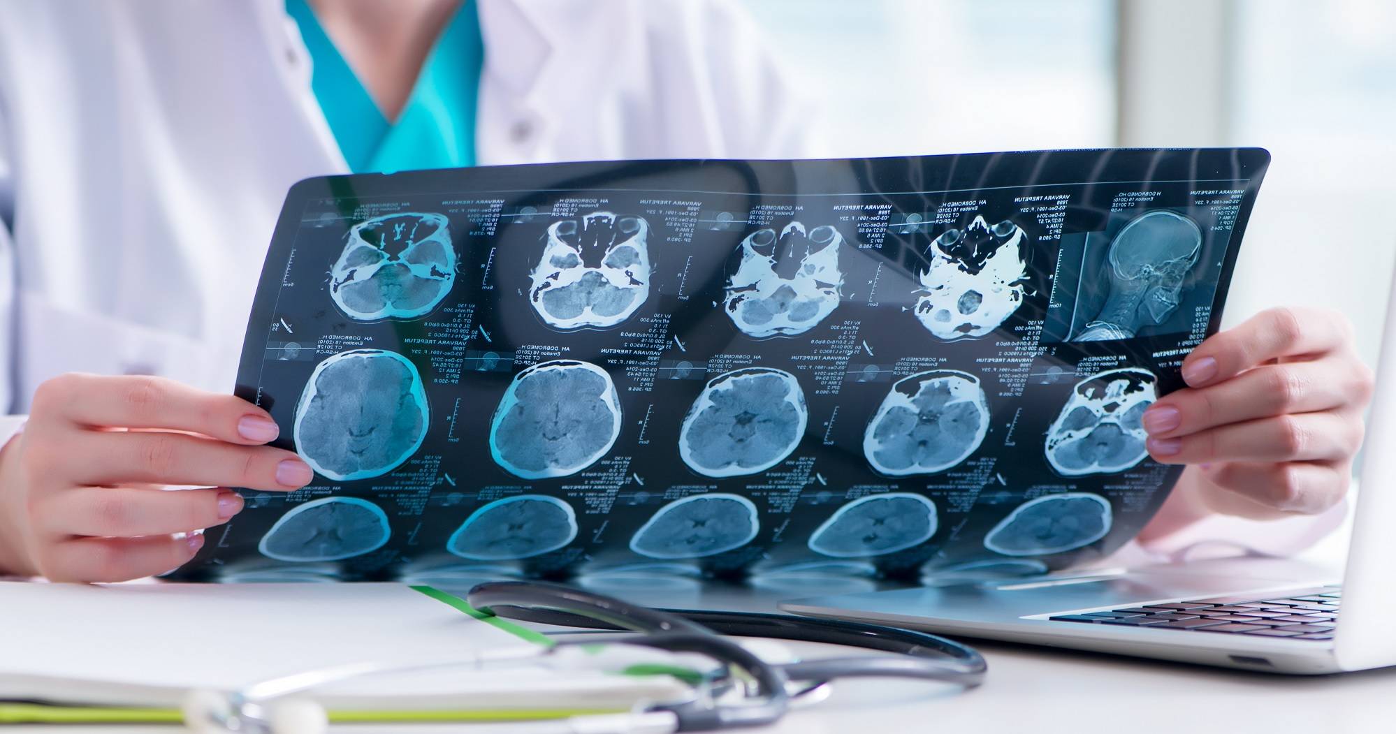
What Is Magnetic Resonance Imaging (MRI) ? 

MRI, which stands for Magnetic Resonance Imaging, is different from X-rays and Computed Tomography (CT) scans in that it is safe and radiation free. The magnetic field produced by an MRI scanner aligns the magnetization of certain atoms in the human body. Radio-frequency pulses are first sent to these atoms which then bounce back, with Nuclear Magnetic Resonance (NMR) signals produced recorded on a computer. The different signals sent by different types of tissues are analyzed and rendered into a detailed image of the internal organs.


Our MRI services include:

  • Cardiac (Heart) MRI scan
    A cardiac MRI generates three-dimensional images of the heart. As a noninvasive and radiation-free technique, it is especially useful for evaluating conditions such as coronary artery disease, heart failure, and congenital heart disease. One single examination is enough to collect detailed information on the heart's pumping and perfusion performance, muscle contractility, blood flow velocity, and condition of the proximal coronary arteries. The test is a convenient, one-stop-shop examination for cardiac patients.
  • Brain MRI scan
    Most specialists agree that the MRI is the best diagnostic tool for the evaluation of the central nervous system (brain and spinal cord). For stroke patients, the diffusion weighted scan allows almost immediate visualization of the parts of the brain with inadequate blood flow, enabling timely treatment to prevent permanent cell death.

  • Spine MRI scan
    A spine MRI provides detailed pictures of areas of the spine which are otherwise hard to see, including the spinal canal, bony segments, and soft tissue. It can pinpoint problems and show the exact location of tumours in the spine, spinal cord, or disks. It is an important diagnostic tool for patients suffering from pain or numbness in the back or the limbs.

  • MRA (Magnetic Resonance Angiography)
    MRA is an examination of the blood vessels including the carotid, pulmonary, peripheral, and renal arteries, as well as the aorta. As a noninvasive technique, MRA also carries less risk for the patient than traditional invasive procedures.

  • Other high-resolution regional MRI scans

    Musculoskeletal and Joint Imaging
    Using MRI to examine muscles and joints from all angles allows for the investigation of complicated soft tissue structure. In addition to providing vital information to orthopaedic surgeons, MRI is also an indispensable tool in the management of sports injuries.


    MR Cholangiogram
    MRI is a noninvasive technique that allows for effective imaging of the entire biliary system, and is most useful in the investigation of the causes of obstruction to the biliary tract.


    Abdominal and Pelvis Imaging
    Breath-hold abdominal and pelvis MRI scans allow for accurate assessments of the conditions of abdominal and pelvic structures.

Computed Tomography Scan

Hong Kong Adventist Hospital – Tsuen Wan is equipped with two Computed Tomography (CT) scanners, the Philips iCT256 Computed Tomography (CT) scanner and the Canon Aquilion ONE/ Prism Edition CT Scanner.


In addition to extremely high speeds, high resolution, and fine details, both scanners also boost lower radiation doses and require less contrast media. The ultrafast scan speeds enable a scan to be completed within seconds, easing the process for patients who may have difficulty holding their breath. The reduced doses of radiation and contrast media also decrease the health risk to patients, especially those with renal diseases who may experience adverse reactions to contrast medium.


Furthermore, the advanced technology of artificial intelligence of the Canon Aquilion ONE/ Prism Edition CT scanner allows the Deep Learning image reconstruction, so that the useful structural images are separated from the useless noises with faster and better image reconstruction.


The Computed Tomography (CT) scan, also called the computerized axial tomography (CAT) scan, is a precise instrument that calculates the degree of X-ray absorbed by human tissues. It produces detailed cross-sectional images of the body parts under examination and compiles them to form three-dimensional images. The short duration of the procedure makes it suitable for children and the elderly, who may have problems lying down for extended periods of time.


Clinical applications of CT scans include:

  • CT Coronary Angiography (CTCA)

    A Computed Tomography Coronary Angiography (CTCA) is a noninvasive and painless screening examination which helps assess a patient’s risk for heart diseases.


    Plaque is formed when fat, calcium, and other substances accumulate in the coronary arteries, gradually narrowing the arteries and blocking blood flow to the heart. This may lead to angina or a heart attack. Our coronary CT scanner provides quick and accurate information about the location and extent of calcified plaques in the coronary arteries.


    Calcium compounds are a major cause of coronary artery disease. The calcium score, which may be computed from data obtained through a CTCA, indicates the degree of calcium blockage in the arteries. Based on the score, suitable treatment for a patient may be designed.


    If you identify with two or more of the following risk factors for coronary heart disease, you should consider a CTCA:

    • Women aged 55 or above or postmenopausal women
    • Men aged 45 or above
    • Family history of heart disease
    • High cholesterol
    • High blood pressure or hypertension
    • Diabetes
    • History of tobacco use
    • Sedentary lifestyle
  • CT Virtual Colonoscopy
    A Virtual Colonoscopy is a recently developed technique that uses a CT scanner and virtual reality software to look inside the large bowel. It is a substitute for conventional colonoscopy, where a long tube is inserted into the colon.

  • CT Angiogram
    A CT angiogram investigates suspected arterial vessel disease without the risk of puncturing blood vessels.

  • Other high-resolution regional CT examinations
  • High-resolution CT for lung diseases
  • Abdominal and pelvic scans
  • Brain scans
Ultrasound Scan (US)
In an ultrasound scan, high-frequency sound waves are transmitted into the patient’s body. The echo sound waves that bounce back are collected with a device, which then displays images of the organ or tissues under examination. The scan is typically used on the gallbladder, the bile ducts, the pancreas, the kidneys, the bladder, the uterus, and the ovaries for detection of conditions such as cirrhosis, gallstones, pancreatic inflammation, and tumours. The absence of radiation in ultrasounds also makes it safe for use in obstetric examinations.
Dual-energy X-ray Absorptiometry (DEXA)

Dual-energy X-ray absorptiometry (DEXA) is the most widely used and most accurate way to measure bone mineral density (BMD). A machine delivers X-ray beams at different energy levels to the spine or hip joints, and its software computes the bone density of the patient by comparing the absorption rates of the two sets of transmissions.


The test is quick and uses very low doses of radiation. It allows patients to keep track of their bone density over the years and is particularly recommended for those with a high risk of osteoporosis, including postmenopausal women, the elderly, and those with a family history of the condition.

Mammography
Mammography

A mammography is a safe and reliable way to evaluate the condition of the breasts. It delivers extremely low doses of X-ray, and can detect breast cancer long before the tumour is large enough to cause symptoms. The US National Cancer Institute (NCI) recommends that women over 40 undergo a mammogram every one to two years. The test is also recommended for younger women with a family history of breast cancer.

Interventional Radiology

The Digital Subtraction Angiography (DSA) machine at our hospital allows for clear visualization of blood vessels in the human body. Digital technology enables artifacts and bony structures that may be blocking the view of the blood vessels to be erased from the image.


The powerful DSA enables interventional radiologists to perform minimally invasive procedures. As technology continues to advance, an increasingly wide range of diseases can now be treated with interventional radiology.

Compared to open surgery, this technique offers a variety of benefits including smaller incisions, less blood loss, and a shorter period of hospitalization, making it a treatment of choice for most patients.

Common interventional radiographic procedures:

  • Uterine fibroid embolization: A proven effective treatment for uterine fibroids
  • Balloon angioplasty/stent: For treatment of peripheral vascular diseases
  • Percutaneous vertebroplasty: A procedure that strengthens vertebral bodies in cases of symptomatic compression fractures caused by osteoporosis or malignancies
  • Thrombolysis: Dissolves blood clots for the treatment of stroke and other thromboembolic diseases
General X-ray and fluoroscopy

X-ray is a type of radiation that is able to pass through different substances at varying absorption rates. With this, images of bones, organs, and other body parts may be recorded and displayed on film, making x-ray tests an excellent method to examine the bones, lungs, and abdominal organs. With the use of contrast media, doctors can also collect images of the urinary system and the digestive system for examination.


The doses of radiation in x-ray tests are generally very low and cause minimal harm to the human body. However, they may cause adverse effects on fetuses and are therefore not suitable for pregnant women.


Our X-ray department provides the following services for both in-patients and out-patients:

  • General X-rays: such as chest and bone X-rays
  • Barium meal: examines the upper alimentary system and stomach
  • Barium enema – examines the colon
  • Intravenous pyelogram or IVP: examines the urinary system for possible obstruction, infection, or mass within the system
  • Hysterosalpingography or HSG: examines the female reproduction system, commonly used for diagnosing infertility
  • Venography: examines the venous system in the body for conditions such as deep vein thrombosis
  • Biopsy for pathology
Positron Emission Tomography and Computed Tomograhy (PET/CT) Scan

The Positron Emission Tomography and Computed Tomography (PET/CT) scan is a dual-modality imaging technique that combines the information of both the metabolic activity and anatomy of the body.  Positron Emission Tomography (PET scan) is an advanced isotope imaging technique that displays the metabolic state of the cell. It can trace and locate the pathological changes of cancer cells in the early stages. Computed Tomography (CT) reveals the structure and location of lesions within the body.


A PET/CT scan is a noninvasive procedure that examines the whole body in one single examination. Any growing tumours may be accurately located, facilitating early detection and treatment of cancer.


Our hospital’s PET/CT scanner is the Philips Ingenuity TF PET/CT, the first PET/CT system to incorporate the image quality improvements of both Astonish TF and iDose.  The system features high-resolution visualization and low dose techniques to manage image quality in both modalities.  The long scan length allows total body studies to be performed in one scan with speed and efficiency.


Clinical applications of PET/CT scan include:

  • Diagnosis and staging of malignancy
  • Evaluation of response and effectiveness of therapy
  • Surveillance of cancer recurrence
Charges for Investigative and Treatment Procedures
Radiological and Imaging Services
Fees and Charges
Charges for Radiological and Imaging Services
Location
199 Tsuen King Circuit, Tsuen Wan, N.T., Hong Kong
Telephone

(852) 2275 6340

Close